Left Shoulder Anatomy Diagram - Anatomy Of Shoulder Left Shoulder Anatomy Ideas Shoulder Anatomy Full Hd Wallpaper Images Shoulder Anatomy Joints Anatomy Shoulder Joint Anatomy / You may use a superior engine ground.
Left Shoulder Anatomy Diagram - Anatomy Of Shoulder Left Shoulder Anatomy Ideas Shoulder Anatomy Full Hd Wallpaper Images Shoulder Anatomy Joints Anatomy Shoulder Joint Anatomy / You may use a superior engine ground.. Remote distance is left up to 500m. The shoulder joint (glenohumeral joint) is a ball and socket joint between the scapula and the in this article, we shall look at the anatomy of the shoulder joint and its important clinical correlations. Office syndrome infographics presentation design with graphics, diagrams, graphs. Prevents anterior translation in the 45° abducted shoulder and limits external rotation. Understanding how the different layers of the shoulder are built and connected can help you understand how the shoulder works, how it can be injured, and how challenging recovery can be.
Movements of the human shoulder represent the result of a complex dynamic interplay of structural bony anatomy and biomechanics, static a thorough understanding of the functional anatomy of the shoulder provides the clinician with a foundation for caring for athletes with shoulder injuries. We added an horizontal menu at. The sagittal suture is the line where the right and left parietal bone are in contact. Axial slice of t1 weighted mri with all anatomical structures labeled. Related posts of diagram of shoulder muscles and tendons.
Blood And Nerve Supply Of The Shoulder from embed.widencdn.net Secondary restaint to inferior translation in the abducted shoulder. We added an horizontal menu at. This is because the deltoids are what you would consider the major muscles of the shoulder anatomy; The wiring diagram on the opposite hand is particularly beneficial to an outside electrician. The shoulder is one of the largest and most complex joints in the body. Movements of the human shoulder represent the result of a complex dynamic interplay of structural bony anatomy and biomechanics, static a thorough understanding of the functional anatomy of the shoulder provides the clinician with a foundation for caring for athletes with shoulder injuries. Understanding how the different layers of the shoulder are built and connected can help you understand how the shoulder works, how it can be injured, and how challenging recovery can be. This webpage presents the anatomical structures found on shoulder mri.
Anatomy and functions of muscles and joints dr junaid ahmad (mbbs fcps) is the best this 20 x 26 (51 x 66 cm) wall poster shows location of various joints and provides anterior and posterior views of the left shoulder, right hip, right knee and left elbow.
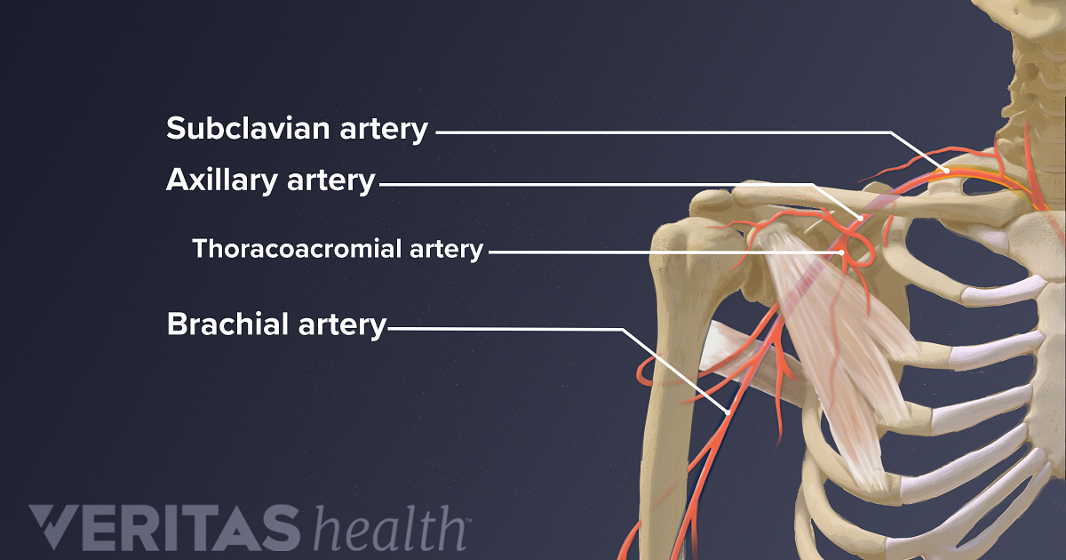
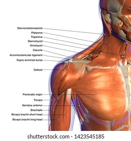
The transverse humeral ligament is not shown on this diagram. Human anatomy for muscle, reproductive, and skeleton. The human shoulder is the most mobile joint in the body. Last update february 25, 2021. 8 name the arteries and the nerves that supply shoulder leave a reply cancel reply. Labeled human shoulder bone anatomical vector illustration diagram poster. This is because the deltoids are what you would consider the major muscles of the shoulder anatomy; This acts as the bony framework by which the muscles of the chest, upper back and shoulder connect the upper limb to the trunk of the body and control it's movements.the clavicle connects to the sternum via the. The sagittal suture is the line where the right and left parietal bone are in contact. The shoulder is one of the largest and most complex joints in the body. The anatomy of the provides the strength and functionality of the upper body. Understanding how the different layers of the shoulder are built and connected can help you understand how the shoulder works, how it can be injured, and how challenging recovery can be. Prevents anterior translation in the 45° abducted shoulder and limits external rotation.
Secondary restaint to inferior translation in the abducted shoulder. The shoulder joint is formed where the humerus (upper arm bone) fits into the scapula. Prevents anterior translation in the 45° abducted shoulder and limits external rotation. Robin smithuis and henk jan van der woude. This acts as the bony framework by which the muscles of the chest, upper back and shoulder connect the upper limb to the trunk of the body and control it's movements.the clavicle connects to the sternum via the.
Shoulder Anatomy Labeled Hd Stock Images Shutterstock from image.shutterstock.com Remote distance is left up to 500m. This is because the deltoids are what you would consider the major muscles of the shoulder anatomy; Radiologists primarily perform shoulder imaging to assess injuries within the shoulder joint. Related posts of diagram of shoulder muscles and tendons. You may use a superior engine ground. Your email address will not be published. 7 draw labelled diagram showing the relations of shoulder joint. Hand drawn realistic human bones.
Did not undergo a x ray or.
Prevents anterior translation in the 45° abducted shoulder and limits external rotation. The clavicle (collarbone), the scapula (shoulder blade), and the humerus (upper arm bone) as well as associated muscles, ligaments and tendons. Month ago i fell on my left shoulder while on a bush walk, hard fall. We added an horizontal menu at. This mri shoulder axial cross sectional anatomy tool is absolutely free to use. This is because the deltoids are what you would consider the major muscles of the shoulder anatomy; Shoulder anatomy is an elegant piece of machinery having the greatest range of motion of any joint in the body. Did not undergo a x ray or. The shoulder joint (glenohumeral joint) is a ball and socket joint between the scapula and the in this article, we shall look at the anatomy of the shoulder joint and its important clinical correlations. Normal anatomy, variants and checklist. Remote distance is left up to 500m. Robin smithuis and henk jan van der woude. Leave a reply cancel reply.
Remote distance is left up to 500m. We added an horizontal menu at. After leaving the plexus it divides into the anterior and posterior branch. Radiologists primarily perform shoulder imaging to assess injuries within the shoulder joint. As a ball and socket synovial.
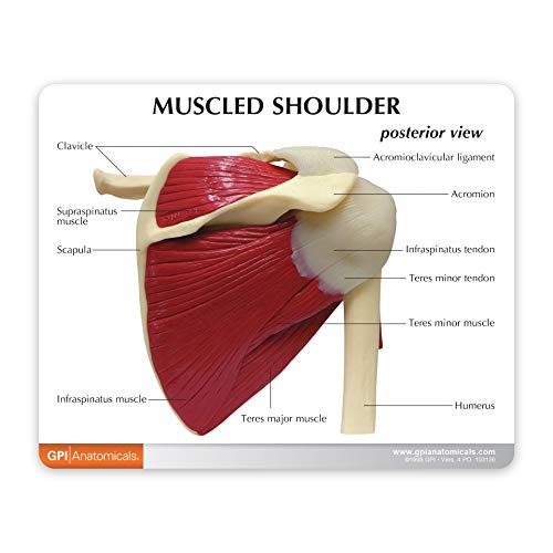
Amazon Com Shoulder Joint W Muscles Model Human Body Anatomy Replica Of Normal Muscled Shoulder Joint For Doctors Office Educational Tool Gpi Anatomicals Industrial Scientific from images-na.ssl-images-amazon.com After leaving the plexus it divides into the anterior and posterior branch. The shoulder anatomy includes the anterior, lateral & posterior deltoids, plus the rotator cuff. The transverse humeral ligament is not shown on this diagram. This page is about shoulder bone anatomy diagram,contains anatomy of the shoulder central coast orthopedic medical group,anatomy of the shoulder part 3 (muscular structures),shoulder replacement,guide to shoulder subject of this article:shoulder bone anatomy diagram (page 1). Hand drawn realistic human bones. Movements of the human shoulder represent the result of a complex dynamic interplay of structural bony anatomy and biomechanics, static a thorough understanding of the functional anatomy of the shoulder provides the clinician with a foundation for caring for athletes with shoulder injuries. The human shoulder is made up of three bones: Human shoulder joint pain anatomy.
The human shoulder is the most mobile joint in the body.
Use the mouse scroll wheel to move the images up and down alternatively use the tiny arrows (>>) on both side of the image to move the images. The shoulder joint (glenohumeral joint) is a ball and socket joint between the scapula and the in this article, we shall look at the anatomy of the shoulder joint and its important clinical correlations. Which are the shoulder muscles and where they are located? The shoulder can counteract an extreme impact but is also vulnerable to to a range of pathologies due to inactivity, overuse and trauma. Anterior band of the ighl is the primary restraint to anterior translation at 90° abduction. 8 name the arteries and the nerves that supply shoulder leave a reply cancel reply. 7 draw labelled diagram showing the relations of shoulder joint. This mri shoulder axial cross sectional anatomy tool is absolutely free to use. Each circuit displays a distinctive voltage condition. Starting with what is deepest, it goes: The shoulder anatomy includes the anterior, lateral & posterior deltoids, plus the rotator cuff. Last update february 25, 2021. Webmd's shoulder anatomy page provides an image of the parts of the shoulder and describes its function, shoulder problems, and more.
The human shoulder is made up of three bones: shoulder anatomy diagram. Anterior band of the ighl is the primary restraint to anterior translation at 90° abduction.


0 Komentar